

Home
About Medical Parasitology
New Infections
Ova & Parasite (O&P) Exams
CPT Codes
Quizzes, General
Quizzes, Histology
Quizzes, Blood
Review Tests
FAQ
Information Tables
Organism Index (A-Z)
A 72-year-old female with a history of congestive heart failure, diabetes, and hypertension was admitted to a local hospital with complaints of progressive shortness of breath. She became hypoxic and was intubated and placed on mechanical ventilation support. X-rays performed showed pulmonary edema. Bronchial alveolar lavage (BAL) specimens were collected and sent to Microbiology and Hematology for routine work-up. During a cell count, one of the technologists noted what appeared to be motile ciliated or flagellated cells. An aliquot of the BAL specimen was also stained with hematoxylin-and-eosin, in which similar objects were observed. (Case reported from Sparks Regional Medical Center, Fort Smith, AR).
What parasitic infection might be involved?
Scroll Down for Answer and Discussion
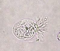
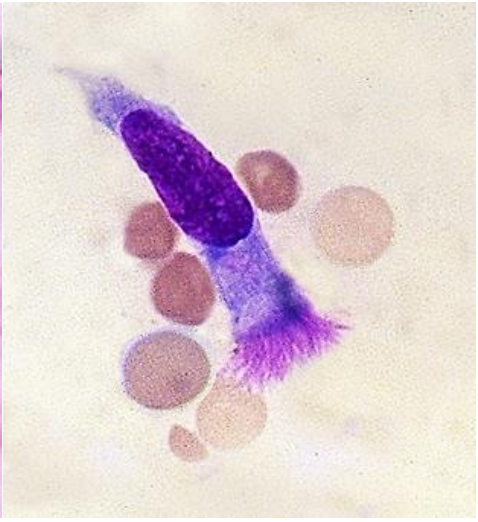
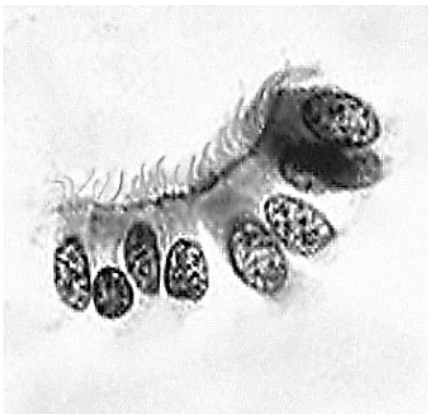
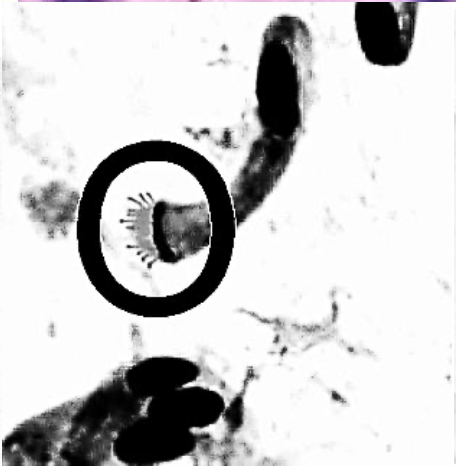
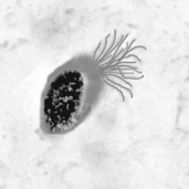
The images presented in Diagnostic Quiz #87 are the following:
The objects shown in this case were detached ciliary tufts (remnants of ciliated epithelial cells) and a diagnosis was made of No Parasites Found. This finding is referred to as ciliocytophthoria, and can be observed in a variety of body fluids, including peritoneal, amniotic, and respiratory specimens. These detached cells are commonly mistaken for protozoa such as Trichomonas, Lophomonas, and Balantidium. There are rare reports documenting Lophomonas in human clinical specimens, and many of them can be attributed to the misidentification of detached epithelial cells.
Detached ciliary tufts (ciliocytophthoria) have been seen in a variety of body fluids (especially peritoneal and amniotic fluids; also respiratory specimens). These tufts are the remnants of ciliated epithelium that are found as a part of normal cellular turnover in a number of sites: respiratory tract and sinuses, ventricles of the brain, central canal of the spinal cord, and epithelia of the male and female reproductive tracts. The tufts are motile, measure 10 to 15 µm in diameter, and can be confused with ciliated or flagellated protozoa.
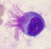
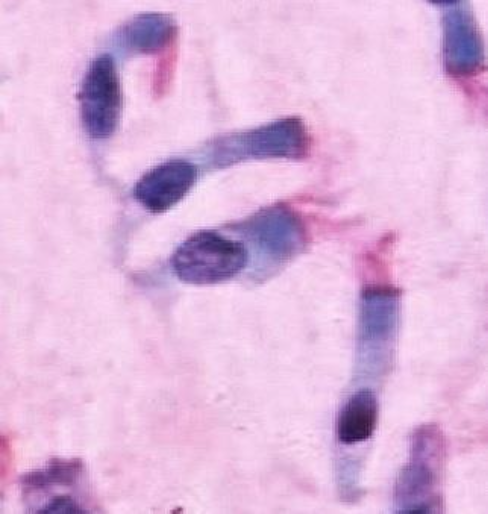
However, when they are carefully examined on a stained smear, there is no internal structure like that seen in protozoa. Ciliated epithelium cells can be seen above; ciliocytophthoria are anucleate remnants of these ciliated cells. However, in some cases, the cell nuclei are clearly visible. The ciliated tufts can be seen on the cells below.
These cells have been found in many clinical specimens, including peritoneal fluid. Recognition of these uncommon structures can be important in avoiding a misdiagnosis with a suspect protozoan infection. This has also been recognized as a potential problem in the virology laboratory. The difference between ciliocytophthoria and ciliated or flagellated parasites becomes very important during examination of specimens submitted for cytologic testing. Also in performing immunofluorescence assays (DFAs) used in the virology laboratory for the rapid detection of viruses, assessment of the cellularity of specimens is required for the most effective use of the DFA assay. Garcia, LS, 2016. Diagnostic Medical Parasitology, 6th Ed., ASM Press, Washington, DC. Each Quiz has a two section format: the first section will present the Quiz topic and the second section will provide a discussion of the answer and/or various options in response to the Quiz situation presented to the user. In some situations, there may be more than one correct response.
The content within this site is made possible through the extensive contribution of Lynne S. Garcia, M.S., MT(ASCP), CLS(NCA), BLM(AAB), F(AAM), Director, Consultantation and Training Services (Diagnostic Medical Parasitology and Health Care Administration). For additional information, she can be contacted at LynneGarcia2@verizon.net. Reference: Garcia, L.S. 2015. Diagnostic Medical Parasitology, 6th Ed., ASM Press, Washington, D.C.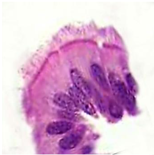
References:
Ashfag-Drewett, R., C. Allen, and R. L. Harrison. 1990. Detached ciliary tufts: comparison with intestinal protozoa and a review of the literature. Am. J. Clin. Pathol. 93:541–545.
Hadziyannis, E., B. Yen-Lieberman, G. Hall, and G. W. Procop. 2000. Ciliocytophthoria in clinical virology. Arch. Pathol. Lab. Med. 124:1220–1223.
Mahoney, C. A., N. Sherwood, E. H. Yap, T. P. Singleton, D. J. Whitney, and P. J. Cornbleet. 1993. Ciliated cell remnants in peritoneal dialysis fluid. Arch. Pathol. Lab. Med. 117:211–213.
Vila, M, K Thompson, and G Erdem. 2011. Motile ciliary microorganisms in peritoneal fluid. Diagn Cytopathol 39:606-607.
Quizzes