Home
About Medical Parasitology
New Infections
Ova & Parasite (O&P) Exams
CPT Codes
Quizzes, General
Quizzes, Histology
Quizzes, Blood
Review Tests
FAQ
Information Tables
Organism Index (A-Z)
***Reminder: Slides are copyrighted and cannot be copied for publication.
A 52 year old male, originally from San Francisco, presented with a three week history of diarrhea and general malaise. This patient was a geologist consultant, and he traveled throughout the world on a regular basis. These trips included Europe, Latin America, Asia, Africa, and throughout North America. He often took side trips on his travels and liked to eat many of the local dishes, since he was an excellent cook. Apparently, he had no underlying health problems. On presentation, he was complaining of intermittent diarrhea, a weight loss of approximately 10 pounds, and general malaise. He had just returned from a trip to Brazil.
Routine stool examinations for parasites (three ova and parasite examinations) revealed the following:

All images photographed using oil immersion (X1,000). Wheatley’s Trichrome Stain
QUESTIONS:
1. What organisms are suggested from the images shown above?
2. What groups of parasites might be involved, and are these typical based on his travel history?
3. What else do these objects suggest?
4. How likely is it that the patient has a mixed infection (based on the structures seen above? Why or why not?
5. What other tests might be requested before all intestinal parasites could be eliminated as possibilities?
(Scroll Down for Answers and Discussion)
ANSWER AND DISCUSSION OF DIAGNOSTIC QUIZ #55
The images presented in Diagnostic Quiz #55 are the following:
ANSWERS TO QUESTIONS (View images from left to right):
1.The structure on the left, photographed using the 100x oil immersion objective (x1,000), resembles a protozoan trophozoite. However, the "nuclei" are not the same size and the morphology is not uniform enough to be protozoan. The clear structures in the next image to the right were photographed using the 100x oil immersion objective (x1,000) and resemble protozoan trophozoites. However, they are not uniform in size and the nucleus takes up a great deal space compared with the cytoplasm. The structures in the next image were also photographed using the 100x oil immersion objective (x1,000) and resemble a protozoan cyst.
2. The structures in the photographs resemble intestinal protozoa. Based on the patient's travel history, any or all of these parasites are possibilities.
3. The images also suggest various artifacts and/or human cells that might be present in human fecal specimens.
4. Based on the images you have seen, it is unlikely that the patient has a mixed infection with multiple parasites. All of the structures are artifacts. However, with the presence of artifacts in the specimen, examination of the stained fecal smears should be very thorough in order to "rule out" the possibility of true parasites.
5. Because routine stool examinations do not "rule out" all the coccidia and/or the microsporidia, additional testing should be performed. Those tests would include fecal immunoassays for Cryptosporidium/Giardia or Cryptosporidium, and special staining for Cyclospora cayetanensis (modified acid-fast stains) and the microsporidia (modified trichrome stains.
COMMENTS ON THE PATIENT:
This case represents a situation in which the cause of the patient's symptoms may be something other than an infection with intestinal parasites. Another possibility is that the routine Ova and Parasite Examination (O&P) did not allow detection and identification of the particular parasite involved. Generally, the coccidia and/or microsporidia require different testing prior to identification (modified acid-fast stains, modified trichrome stains).
COMMENTS ON THE METHOD LIMITATIONS:
It is always recommended that a thorough examination be performed (minimum of two specimens for O&P examinations - three is even better) before reporting the patient as "No Parasites Seen." Also, remember that the routine O&P examination does not allow the detection of organisms like Cryptosporidium spp., Cyclospora cayetanensis, or the microsporidia. A statement listing the method limitations should be included in the report to the physician. In spite of these method limitations with some of the coccidia, the coccidian, Isospora belli, can usually be identified in the concentration sediment wet smear examination.
Wheatley's trichrome stain (permanent stained smear) (x1,000)
|
|
|
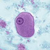
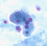
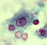
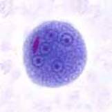
Macrophage Entamoeba histolytica/E. dispar Entamoeba histolytica
Although this structure on the left is the same size as an Entamoeba histolytica/E. dispar trophozoite, the nucleus takes up more space than one in a true protozoan trophozoite. This cell is a macrophage (human cell) and in the lower right, a polymorphonuclear leukocyte (PMN). In comparison, the image in the middle is a true protozoan trophozoite, Entamoeba histolytica/E. dispar. Notice the different ratios between the nuclear material and cytoplasm. This is one of the critical ways to differentiate human cells from actual protozoa. Also, note the image on the right containing ingested RBCs in the cytoplasm – this specific morphology confirms the organism is the true pathogen, Entamoeba histolytica.
Wheatley's trichrome stain (permanent stained smear) (x1,000)
Although these structures (wet mount) tend to resemble protozoa, notice that the sizes are somewhat different and the overall shape/size are not consistent. These are PMNs. With relatively fresh blood in the stool, the PMNs often resemble the same cells that are seen in a routine hematology smear (lobed nuclei). However, if the blood has been in the stool for some time, the lobed nuclei break apart and tend to resemble protozoan cysts.
Wheatley's trichrome stain (permanent stained smear) (x1,000)
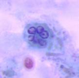
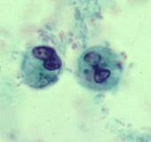
PMN Entamoeba coli cyst
Although this structures (permanent stained smear) tends to resemble a protozoan cyst, notice that the cell nuclei tend to take up a very large part of the cell; this large ratio of nuclear material to cytoplasm (about 1:2) is much more typical of WBCs, not intestinal protozoa. This cell represents a PMN that has been in the gut for a while. Notice that the lobed nucleus has come apart and the resulting structure can mimic a protozoan cyst. Notice the protozoan cyst on the right (less nuclear material per cytoplasm than is seen in the PMN).
REFERENCES
Garcia, LS, 2016. Diagnostic Medical Parasitology, 6th Ed., ASM Press, Washington, DC.
Leber, A.L. and S.M. Novak. 2003. Intestinal and Urogenital Amebae, Flagellates, and Ciliates In: Murray, P.R., E.J. Baron, J.H. Jorgensen, M.A. Pfaller, and R.H. Yolken (eds). Manual of Clinical Microbiology, 8th ed, vol 2, ASM Press, Washington, D.C.
Each Quiz has a two section format: the first section will present the Quiz topic and the second section will provide a discussion of the answer and/or various options in response to the Quiz situation presented to the user. In some situations, there may be more than one correct response.
The content within this site is made possible through the extensive contribution of Lynne S. Garcia, M.S., MT(ASCP), CLS(NCA), BLM(AAB), F(AAM), Director, Consultantation and Training Services (Diagnostic Medical Parasitology and Health Care Administration). For additional information, she can be contacted at LynneGarcia2@verizon.net.
Reference: Garcia, L.S. 2015. Diagnostic Medical Parasitology, 6th Ed., ASM Press, Washington, D.C.