Home
About Medical Parasitology
New Infections
Ova & Parasite (O&P) Exams
CPT Codes
Quizzes, General
Quizzes, Histology
Quizzes, Blood
Review Tests
FAQ
Information Tables
Organism Index (A-Z)
A patient is a 48 year-old male from Japan who has traveled throughout the world as a professional photographer (Europe, Asia, Central and South America, Australia, New Zealand, etc.). He was diagnosed as having diarrhea, cough, and general malaise. He had a history of vague health problems over the past few years, but was never sick enough to see a physician. Three stool specimens were submitted to the laboratory. After examination of the concentration sediment and permanent stained smears from all three specimens, the following objects were seen. Please comment on the identification of the structures seen.
1. 60 x 40 µm |
2. 30 x 15 µm |
3. 140 x 70 µm |
4. 90 x 50 µm |
5. 360 x 20 µm |
6. 18 µm |
Scroll Down for Answer and Discussion
Answer and Discussion of Quiz #1
The images presented in Diagnostic Quiz #4 are the following:
Comments on the patient:
The patient may have experienced vague abdominal symptoms from several of the parasites found, including Isospora, hookworm or Fasciolopsis buski/Fasciola hepatica. The cough is probably related to the Paragonimus spp. infection, often causing symptoms on a random basis (particularly when egg production leads to cough and the production of sputum containing the egg packets). When Paragonimus spp. eggs are found in the stool, the sputum can also be checked; however, specimens may be negative unless the patient is symptomatic at the time. Isospora belli infections are not that common and are usually associated with symptoms in the immunocompromised patient; in this case, it may be an incidental finding. Symptoms with either F. buski or F. hepatica are definitely related to parasite burden; a light infection will generally cause no symptoms. Entamoeba coli is a nonpathogen and would merely be an indication that the patient had ingested something contaminated with fecal material.
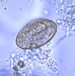
Hookworm:
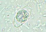
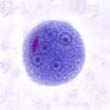
Isospora belli:
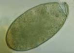
Fasciolopsis buski:
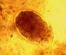
Fasciola hepatica:
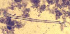
Paragonimus spp.:
Artifact - Root hair:
Plant or root hairs, such as the fuzz on peaches, may resemble nematode larvae. The root hairs tend to be clear and refractile while the larvae pick up stain (iodine), which will reveal internal structures. It is important to recognize this potential error when examining formalin-fixed specimens submitted as proficiency testing specimens. In cases of diarrhea, partially digested plant material, examples being bean sprouts or other vegetable material, can mimic adult nematodes or tapeworm proglottids. Also, all stages of free-living nematodes can occur in feces or as contaminants of water used in making fecal suspensions.
Entamoeba coli:
With the exception of the mature cyst, the morphologies of E. histolytica and E. coli are similar in most of the stages. Therefore, it is very important to examine permanent stained smears, even if a tentative identification has been made from a wet preparation examination. Specific treatment is not recommended for this nonpathogen. Consequently, the correct differentiation between the two species is critical to good patient care. Since the two species are acquired in the same way, it is also important to remember that both can be found in the same patient. If few E. histolytica are present among many E. coli organisms, additional searching may be necessary to correctly identify both. Remember: Entamoeba histolytica can be diagnosed using morphologic criteria ONLY if RBCs are seen within the cytoplasm of the trophozoite. Otherwise, the organisms should be identified as Entamoeba histolytica/E. dispar.
Review (Consistent with CAP Inspection Checklist):
Every stool submitted for an O&P examination must be examined using the concentration and permanent stained smear procedures.
O&P Exam (Fresh Stool Specimen/liquid and/or very soft): Direct wet smear, concentration, permanent stained smear.
O&P Exam (Fresh Stool Specimen/formed stool): Concentration, permanent stained smear. O&P Exam (Preserved Stool Specimen): Concentration, permanent stained smear. Remember that all intestinal protozoan infections can be missed if the concentration ONLY is performed. The permanent stained smear is much more sensitive than the concentration alone.
To date, there are no commercial immunoassay products available for the confirmation of infection with D. fragilis.
Additional Reading:
Each Quiz has a two section format: the first section will present the Quiz topic and the second section will provide a discussion of the answer and/or various options in response to the Quiz situation presented to the user. In some situations, there may be more than one correct response.
The content within this site is made possible through the extensive contribution of Lynne S. Garcia, M.S., MT(ASCP), CLS(NCA), BLM(AAB), F(AAM), Director, Consultantation and Training Services (Diagnostic Medical Parasitology and Health Care Administration). For additional information, she can be contacted at LynneGarcia2@verizon.net.
Reference: Garcia, L.S. 2015. Diagnostic Medical Parasitology, 6th Ed., ASM Press, Washington, D.C.