

Home
About Medical Parasitology
New Infections
Ova & Parasite (O&P) Exams
CPT Codes
Quizzes, General
Quizzes, Histology
Quizzes, Blood
Review Tests
FAQ
Information Tables
Organism Index (A-Z)
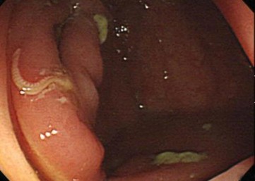
A 57 year old male presented for a routine screening colonoscopy after complaining of minor, vague lower abdominal discomfort. A series of routine O&P examinations was negative. During colonoscopy, he was found to have a small, white worm on the proximal ascending colon. One end appeared to be embedded in the colonic mucosa, while the other end was coiled and motile. The following image was seen:
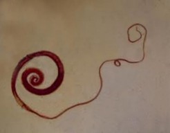
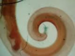
Using forceps, the parasite was removed and examined microscopically. The following images were seen.
What organisms are suggested from the images shown above?
What groups of parasites might be involved, and are these typical based on his history?
If this is a helminth, why was the O&P examination negative?
How common is this parasite worldwide and how likely is it that the patient might be asymptomatic?
The images certainly suggest a helminth; the overall appearance is that of a roundworm (nematode).
The presence of a nematode infection is certainly a possibility, particularly since many patients may be asymptomatic or present with minor symptoms.
Because this helminth was diagnosed as a male Trichuris trichiura (whipworm) and no female worms were seen, it is not uncommon to have a negative O&P examination with no eggs seen in the stool specimen.
This infection is very common worldwide, particularly in the tropics and subtropics. It is often found as a mixed infection with Ascaris lumbricoides, since both helminths are acquired from the ingestion of mature eggs from contaminated soil or water.
COMMENTS ON THE PATIENT:
Trichuriasis is an intestinal infection caused by the ingestion of embryonated eggs from the environment. Colonized eggs hatch and enter the crypts of the small intestine as larvae. After 1-3 months of maturation, the parasite migrates to the cecum. In the cecum, the parasite matures, mates, and lays eggs. Adult worms are 3-4 cm in length and have thin, tapered anterior regions, and are thus commonly referred to as whipworms. The adult T. trichiura invade the mucosa and produce minor inflammatory changes at localized sites. In endemic areas, most people are colonized by small numbers of worms and have no symptoms. The head end of the nematode embeds in the colonic mucosa, while the tail end is free in the lumen.
COMMENTS ON THE METHOD RECOMMENDATIONS:
Although the O&P examinations were negative, this is not uncommon with the presence of a single male adult worm. No eggs would be expected in the stool specimen without the presence of a female worm, as well. The laboratory findings were normal without definite eosinophilia (very light infection). On colonoscopy, parasites have been incidentally reported in the cecum and in the sigmoid colon.
COMMENTS ON THE IMAGES:
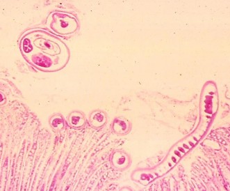
The images are very typical. Note the curved tail of the male worm. Per histology, the following image is very typical with the head end embedded in the mucosa while the tail end is free in the lumen (see green arrow). Note the larger structures free in the lumen (body of the worm), while the smaller head end is embedded (see black arrow). The head end (whip portion) is much thinner than the body portion (handle) as seen below.
Garcia, L.S. 2016. Diagnostic Medical Parasitology, 6th Ed., ASM Press, Washington, D.C.
Each Quiz has a two section format: the first section will present the Quiz topic and the second section will provide a discussion of the answer and/or various options in response to the Quiz situation presented to the user. In some situations, there may be more than one correct response.
The content within this site is made possible through the extensive contribution of Lynne S. Garcia, M.S., MT(ASCP), CLS(NCA), BLM(AAB), F(AAM), Director, Consultantation and Training Services (Diagnostic Medical Parasitology and Health Care Administration). For additional information, she can be contacted at LynneGarcia2@verizon.net.
Reference: Garcia, L.S. 2015. Diagnostic Medical Parasitology, 6th Ed., ASM Press, Washington, D.C.