Home
About Medical Parasitology
New Infections
Ova & Parasite (O&P) Exams
CPT Codes
Quizzes, General
Quizzes, Histology
Quizzes, Blood
Review Tests
FAQ
Information Tables
Organism Index (A-Z)
A 33 year old female, originally from Mexico, was admitted to the hospital with seizures. This patient had previously experienced convulsions when she was 14, but had remained asymptomatic until about a year prior to admission. The seizures had become more numerous and now occurred about once a month. The patient had experienced an inability to talk and had become unconscious for several minutes during these episodes. She had also complained of double vision.
On admission, the patient appeared to have partial paralysis on the right side and complained of progressive weakness, again on the right side. Computed tomographic (CT) and magnetic resonance imaging (MRI) studies revealed several intracerebral lesions, some of which appeared to be calcified. On surgery, the following structures were seen and removed:
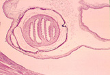
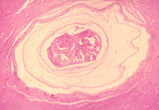
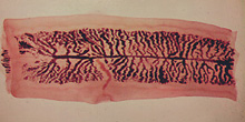
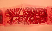
Gross specimen, measuring 3 by 4 cm.
Three cysts were removed. Routine pathology preparations revealed the following:
|
|
Images courtesy of A Pictorial Presentation of Parasites: A cooperative collection prepared and/or edited by H. Zaiman)
Scroll Down for Answer and Discussion
Answer and Discussion of Quiz #29
The images presented in Diagnostic Quiz #29 are the following:
Answers to Questions:
Comments on the Patient: This case represents an infection with Taenia solium, the cause of cysticercosis. When these larvae are found in the brain, symptoms can result from actual larval invasion of the brain tissue and/or death or the organism, which stimulates tissue reactions the larvae. It has been recommended that in every case of epilepsy occurring in a patient with no family history of seizures and no previous history of seizures in childhood, the probability of cysticercosis should be considered. Other symptoms, including abnormal behavior, transient paresis, intermittent obstructive hydrocephalus, disequilibrium meningoencephalitis, and visual problems, may also be present. In this patient there was a history of seizures in childhood, and many of the key symptoms of cysticercosis were also present.
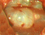
Comments on the Organism: The cysticercus is an ovoid, milky white bladder with the head invaginated into the bladder. The bladder worms measure 5 mm long by 8 to 10 mm wide, except in brain or subminingeal spaces where they can have a volume of about 60 ml. The larval forms of T. solium (cysticerci) have been recovered from all areas of the body, and symptoms depend on the body site involved. The presence of cysticerci in the brain represents the most frequent parasitic infection of the human nervous system and the most common cause of adult-onset epilepsy throughout the world.
On ingestion by hogs or humans, the eggs of T. solium hatch in the duodenum or jejunum after exposure to gastric juice in the stomach. The released oncospheres penetrate the intestinal wall, are carried via the mesenteric venules throughout the body, and are filtered out in the subcutaneous and intramuscular tissues, the eyes, the brain, and other body sites. Neurocysticercosis usually affects males and females of all ages, with a peak incidence between 30 and 50 years of age.
Taenia solium Key Points - Laboratory Diagnosis
ADULT WORM
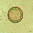
CYSTICERCI
|
|
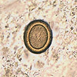
Taenia spp. eggs containing six-hooked oncosphere: note the striated shell
|
|
Taenia saginata (beef tapeworm) and Taenia solium (pork tapeworm) gravid proglottids: note the number of branches
Each Quiz has a two section format: the first section will present the Quiz topic and the second section will provide a discussion of the answer and/or various options in response to the Quiz situation presented to the user. In some situations, there may be more than one correct response.
The content within this site is made possible through the extensive contribution of Lynne S. Garcia, M.S., MT(ASCP), CLS(NCA), BLM(AAB), F(AAM), Director, Consultantation and Training Services (Diagnostic Medical Parasitology and Health Care Administration). For additional information, she can be contacted at LynneGarcia2@verizon.net.
Reference: Garcia, L.S. 2015. Diagnostic Medical Parasitology, 6th Ed., ASM Press, Washington, D.C.