Presentation of Quiz #52
A patient is a 52 year-old male from the United States who has traveled throughout the world as a professional consulting engineer (Europe, Asia, Central and South America, Australia, New Zealand, etc.). He was complaining of diarrhea, cough, abdominal discomfort, and general malaise. He has no serious underlying health problems, and was not currently under a physician's care. He presented to his regular physician and, subsequently, three stool specimens were submitted to the laboratory. After examination of the concentration sediment and permanent stained smears from all three specimens, the following objects were seen. Please comment on the identification of the structures seen.
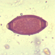
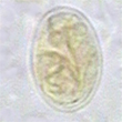
1: High Dry Objective, 2: High Dry Objective (enlarged), 3: Oil Immersion Objective (>12 µ)
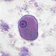
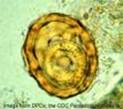
4: High Dry Objective, 5: Oil Immersion Objective, 6: Oil Immersion Objective
Scroll Down for Answer and Discussion
Answer and Discussion of Quiz #52
The images presented in Diagnostic Quiz #52 are the following:
- Trichuris trichiura egg
- Giardia lamblia cyst
- Entamoeba histolytica/E. dispar trophozoite
- Hymenolepis nana egg
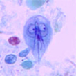
- Giardia lamblia trophozoite
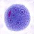
- Entamoeba coli cyst
Comments on the Patient:
The patient may have experienced vague abdominal symptoms from several of the parasites found, including Trichuris trichiura, Ascaris lumbricoides, or Giardia lamblia. The cough may be related to other causes. Entamoeba coli is a nonpathogen and would merely be an indication that the patient had ingested something contaminated with fecal material. The morphology of Entamoeba histolytica/E. dispar does not allow differentiation between the true pathogen E. histolytica and the nonpathogen E. dispar; without the use of species-specific immunoassays. STOOL TESTING RECOMMENDED ORDERS
Trichuris trichiura:
- Eggs are normally passed in the stool and the typical morphology (barrel-shaped with two polar plugs, one at each end of the egg) leads to the correct identification. Even on the permanent stained smear, the typical morphology is usually visible, while many other helminth eggs tend to be distorted on the permanent stained smear.
- These eggs are not infective when passed, but require a period of time in the soil, during which the egg develops.
- Infections with T. trichiura are common worldwide and the infection is acquired through ingestion of infective eggs from contaminated food and/or water.
Giardia lamblia:
- The cysts are easily seen in the concentration wet mount, using the low power objective. More detail can be seen using the high dry objective. Definitive identification is usually confirmed from the permanent stained smear.
- This infection is very common worldwide; patients may range from being asymptomatic to having symptoms of a malabsorption syndrome with frequent diarrhea, cramps, and gas.
- Because the organisms tend to adhere to the mucosal lining, the infection can be difficult to diagnose; multiple stool examinations may be required. It is also important to remember that fecal immunoassays may not always be positive in situations where the number of organisms present is low; the immunoassay might have to be repeated.
- Multiple Ova and Parasite examinations may also be negative in patients with a very light infection; also the organisms are shed on a periodic basis. Not all three stool specimens are likely to be positive.
Entamoeba histolytica/E. dispar:
- These organisms measure >12 microns in the trophozoite form; if the measurement was <12 microns, the trophozoite would be identified as Entamoeba hartmanni.
- Since the trophozoites do not contain ingested RBCs, one cannot identify the organism as the true pathogen, Entamoeba histolytica. However, there are fecal immunoassays available that can confirm the presence of the E. histolytica/E. dispar group or the true pathogen, E. histolytica. These are EIA procedures and require fresh and/or frozen stool specimens for testing; formalinized specimens or those preserved in PVA are not acceptable.
- Based on the organism morphology seen in the permanent stained smear, it is not possible to tell whether the true pathogen, E. histolytica, is present.
Ascaris lumbricoides:
- This typical roundworm egg is characterized by the somewhat round/oval shape and the presence of a very bumpy (tuberculated) egg shell. This egg is fertilized; if unfertilized, the egg would be somewhat larger, more oval, and the bumpy coat would also be prominent. Occasionally, eggs are seen (both fertilized and unfertilized) that have lost the bumpy egg shell; these are called decorticate eggs. Note this egg shell contains a larval worm (eggs have been held for some time – photograph taken from fixed specimen – most fixatives will not kill Ascaris eggs.
- This nematode life cycle is somewhat different in that after egg ingestion from the soil (similar to that seen with T. trichiura eggs), there is an extensive migratory pathway through the heart, lungs, trachea, and the adult worms then develop to maturity within the GI tract. It is not uncommon to find dual infections with both Ascaris and Trichuris, since both are acquired from the ingestion of eggs from contaminated soil.
- Patient symptoms are based on the number of eggs ingested; low numbers may not cause any symptoms, while high egg numbers may lead to symptoms during the migratory pathway within the life cycle.
- The wet preparation can be examined using the 10X (low power) objective, these eggs are large enough that they can be seen using this magnification. Additional morphology could be seen using the high dry objective. It is important not to use too much iodine; if too much iodine is present, the eggs may appear very dark and look like debris.
- On a permanent stained smear, these eggs tend to appear somewhat shrunk and distorted; the eggs may resemble stool debris.
Entamoeba coli:
- With the exception of the mature cyst, the morphologies of E. histolytica and E. coli are similar in most of the stages. Therefore, it is very important to examine permanent stained smears, even if a tentative identification has been made from a wet preparation examination. In the typical cyst, if five or more nuclei can be seen, the organism can be identified as E. coli.
- Specific treatment is not recommended for this nonpathogen. Consequently, the correct differentiation between the two species (E. coli vs. E. histolytica/E. dispar is critical to good patient care.
- Since the three species are acquired in the same way, it is also important to remember that all three can be found in the same patient.
- If few E. histolytica/E. dispar are present among many E. coli organisms, additional searching may be necessary to correctly identify both.
Remember:
Entamoeba histolytica can be diagnosed using morphologic criteria ONLY if RBCs are seen within the cytoplasm of the trophozoite. Otherwise, the organisms should be identified as E. histolytica/E. dispar.
Review (Consistent with CAP Inspection Checklist): Every stool submitted for an O&P examination must be examined using the concentration and permanent stained smear procedures.
O&P Exam (Fresh Stool Specimen/liquid and/or very soft): Direct wet smear, concentration, permanent stained smear.
O&P Exam (Fresh Stool Specimen/formed stool): Concentration, permanent stained smear. O&P Exam (Preserved Stool Specimen): Concentration, permanent stained smear. Remember that all intestinal protozoan infections can be missed if the concentration ONLY is performed. The permanent stained smear is much more sensitive than the concentration alone.
Additional Reading:
- Garcia, LS, 2016. Diagnostic Medical Parasitology, 6th Ed., ASM Press, Washington, DC.
- Garcia, LS, 2009. Practical Guide to Diagnostic Parasitology, 2nd Ed., ASM Press, Washington, DC
- NCCLS, 1997, Procedures for the recovery and identification of parasites from the intestinal tract, Approved Guideline, M28-A, National Committee for Clinical Laboratory Standards, Villanova, PA.
Quizzes
Each Quiz has a two section format: the first section will present the Quiz topic and the second section will provide a discussion of the answer and/or various options in response to the Quiz situation presented to the user. In some situations, there may be more than one correct response.
The content within this site is made possible through the extensive contribution of Lynne S. Garcia, M.S., MT(ASCP), CLS(NCA), BLM(AAB), F(AAM), Director, Consultantation and Training Services (Diagnostic Medical Parasitology and Health Care Administration). For additional information, she can be contacted at LynneGarcia2@verizon.net.
Reference: Garcia, L.S. 2015. Diagnostic Medical Parasitology, 6th Ed., ASM Press, Washington, D.C.