Home
About Medical Parasitology
New Infections
Ova & Parasite (O&P) Exams
CPT Codes
Quizzes, General
Quizzes, Histology
Quizzes, Blood
Review Tests
FAQ
Information Tables
Organism Index (A-Z)
A 4-year-old boy was taken to see his pediatrician with complaints of diarrhea and failure to gain weight. The child weighed 6 lbs. 8 oz. at birth and appeared normal. Developmentally, he was on track for his age. About 18 months before, his family had spent six months in various locations in South America. Following that stay outside of the United States, the mother indicated the child had continued to have intermittent diarrhea, had not gained weight, and was sometimes irritable or listless. Occasionally, she had seen blood in his stool. She also described on one occasion, "His insides were protruding out of his rectum."
The examination revealed a normally proportioned child who weighed 40 lbs., has somewhat shrunken eyes, and appeared unhappy. A partially prolapsed rectum was seen with white objects on the mucosa. Other than these findings, the examination was normal. Stool examinations revealed the following:
Answer and Discussion of Quiz #17
The image presented in Diagnostic Quiz #17 is the following:
Comment: Light infections may not be treated; patients may be asymptomatic, with eggs an incidental finding. However, in this case, the patient had a more severe infection with occasional dysentery and rectal prolapse. The dysentery seen in severe infections may mimic that seen in amebiasis. Worms tend to concentrate in the cecum, but small numbers may localize in any portion of the colon, including the appendix. The adult worm embeds its narrow anterior end into the intestinal mucosa. Host tissue response is usually mild, with local chronic inflammation and eosinophilia. Charcot-Leyden crystals can be found in the stool, and a mild to moderate peripheral eosinophilia is common.
Mebendazole is currently the drug of choice. However, caution must be taken when giving mebendazole to patients with multiple helminth infections, since mebendazole can stimulate erratic migration of Ascaris, with serious consequences.
Most whipworm infections can be easily diagnosed by finding the characteristic eggs in the stool. These eggs should be quantitated (rare, few, moderate, many), since light infections usually cause no problems and may not require therapy.
 |
|
|
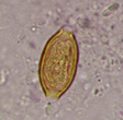
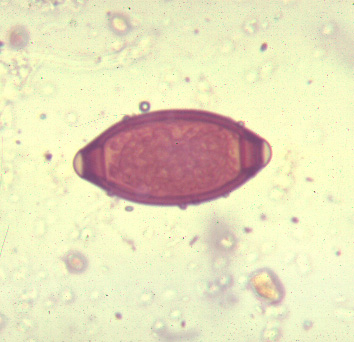
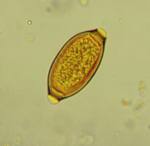
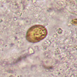
Three images of Trichuris trichiura eggs from a routine stool examination. The first two images are taken from wet preparations. The far left image shows an egg turned on end; this presentation may be difficult to identify as a T. trichiura egg. The middle image shows an egg photographed in a routine saline wet preparation from a concentration sediment. The image on the far right represents an egg seen in a trichrome permanent stained smear.
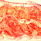
This is an image showing the adult whipworms attached to the intestinal mucosa in a severe case of infection with T. trichiura. Note the white worms.
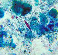
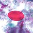
This is an image showing Charcot-Leyden crystals in the stool; this is a trichrome permanent stained smear and the crystal stains red and has two pointed ends. These crystals are formed from the breakdown products of eosinophils and indicate an immune response. However, the presence of these crystals may not always be attributed to a parasitic infection.
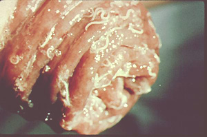
This is an image demonstrating rectal prolapse. Note the white adult worms seen on the surface of the mucosa.
References:
Each Quiz has a two section format: the first section will present the Quiz topic and the second section will provide a discussion of the answer and/or various options in response to the Quiz situation presented to the user. In some situations, there may be more than one correct response.
The content within this site is made possible through the extensive contribution of Lynne S. Garcia, M.S., MT(ASCP), CLS(NCA), BLM(AAB), F(AAM), Director, Consultantation and Training Services (Diagnostic Medical Parasitology and Health Care Administration). For additional information, she can be contacted at LynneGarcia2@verizon.net.
Reference: Garcia, L.S. 2015. Diagnostic Medical Parasitology, 6th Ed., ASM Press, Washington, D.C.