

Home
About Medical Parasitology
New Infections
Ova & Parasite (O&P) Exams
CPT Codes
Quizzes, General
Quizzes, Histology
Quizzes, Blood
Review Tests
FAQ
Information Tables
Organism Index (A-Z)
***Reminder: Slides are copyrighted and cannot be copied for publication.
A 53 year old male, originally from Los Angeles, was seen in the clinic with a three week history of diarrhea and general malaise. This patient was a technical equipment buyer for his company, and he traveled throughout the world visiting suppliers on a regular basis. These trips included Europe, Latin America, Asia, Africa, and throughout North America. He often took side trips on his travels and tended to stay in both expensive and relatively inexpensive accommodations. He also liked to eat many of the local dishes on his travels. Apparently, he had no underlying health problems. On presentation to the clinic, he was complaining of intermittent diarrhea, a weight loss of approximately 10 pounds, and general malaise.
Routine stool examinations for parasites (three ova and parasite examinations) revealed the following:
 |
 |
 |
QUESTIONS:
1. What organisms are suggested from the images shown above?
2. What groups of parasites might be involved, and are these typical based on his travel history?
3. What else do these objects suggest?
4. How likely is it that the patient has a mixed infection (based on the structures seen above? Why or why not?
5. What other tests might be requested before all intestinal parasites could be eliminated as possibilities?
(Scroll Down for Answers and Discussion)
ANSWER AND DISCUSSION OF DIAGNOSTIC QUIZ #62
ANSWERS TO QUESTIONS:
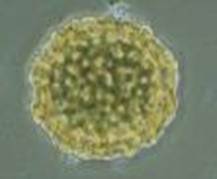
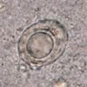
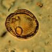
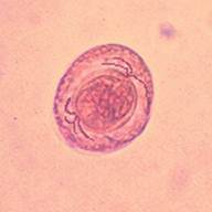
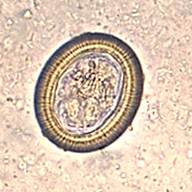
1. The yellow structure, photographed using the 40x high dry objective (x400), resembles a helminth egg, possibly Ascaris lumbricoides. However, the "egg" is too round and the bumps on the egg shell are not pronounced enough for a fertilized egg. Also, the color is not right for an Ascaris egg; they tend to be more tan to brown in a fecal wet mount. The clear structure in the center image was photographed using the 40x high dry objective (x400) and also resembles a helminth egg, particularly Hymenolepis nana. However, there is no internal structure (six-hooked oncosphere) or polar filaments. The structure in the right image was photographed using the 40x high dry objective (x400) and resembles a different type of helminth egg, possibly a Taenia spp. egg. However, the egg is neither round nor oval and there is no striated shell.
2. The structures in the photographs resemble intestinal helminth eggs. Based on the patient's travel history, any or all of these parasites are possibilities.
3. The images also suggest various artifacts that might be present in human fecal specimens.
4. Based on the images you have seen, it is unlikely that the patient has a mixed infection with multiple parasites. All of the structures are artifacts, probably pollen grains. However, with the presence of numerous artifacts in the specimen, examination of the stained fecal smears should be very thorough in order to "rule out" the possibility of true parasites, possibly intestinal protozoa.
5. Because routine stool examinations do not "rule out" all the coccidia and/or the microsporidia, additional testing should be performed. Those tests would include immunoassays for Cryptosporidium/Giardia or Cryptosporidium, and special staining for Cyclospora cayetanensis (modified acid-fast stains) and the microsporidia (modified trichrome stains.
STOOL TESTING RECOMMENDED ORDERS
COMMENTS ON THE PATIENT:
This case represents a situation in which the cause of the patient's symptoms may be something other than an infection with intestinal parasites. Another possibility is that the routine Ova and Parasite Examination (O&P) did not allow detection and identification of the particular parasite involved, particularly if the patient is infected with coccidia and/or microsporidia. Based on the patient history of travel and food preferences, it is always possible the patient ingested something that contained infective parasites. However, in this case, the cause of his symptoms may not be due to an infectious agent.
COMMENTS ON THE METHOD LIMITATIONS:
It is always recommended that a thorough examination be performed (minimum of two specimens for O&P examinations - three is even better) before reporting the patient as "No Parasites Seen." Also, remember that the routine O&P examination does not allow the detection of organisms like Cryptosporidium spp., Cyclospora cayetanensis, or the microsporidia. A statement listing the method limitations should be included in the report to the physician. In spite of these method limitations with some of the coccidia, the coccidian, Isospora belli, can usually be identified in the concentration sediment wet smear examination.
COMMENTS ON ACTUAL PARASITE IMAGES:
 |
 |
 |
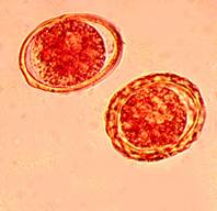
The image on the left represents two Ascaris lumbricoides eggs; note the decorticate egg shell in the left image and the typical bumpy egg shell on the right. The middle image represents a Hymenolepis nana egg; note the suggestion of hooklets within the oncosphere (larva) and the stringy polar filaments that lie between the oncosphere and the egg shell. The image on the right is a Taenia spp. egg; note the hooklets within the egg shell and the typical striations on the egg shell. If these images are compared with the artifact images, it is obvious that the artifacts do not have the key characteristics necessary to identify the objects as actual parasites.
REFERENCES
Garcia, LS, 2016. Diagnostic Medical Parasitology, 6th Ed., ASM Press, Washington, DC.
Each Quiz has a two section format: the first section will present the Quiz topic and the second section will provide a discussion of the answer and/or various options in response to the Quiz situation presented to the user. In some situations, there may be more than one correct response.
The content within this site is made possible through the extensive contribution of Lynne S. Garcia, M.S., MT(ASCP), CLS(NCA), BLM(AAB), F(AAM), Director, Consultantation and Training Services (Diagnostic Medical Parasitology and Health Care Administration). For additional information, she can be contacted at LynneGarcia2@verizon.net.
Reference: Garcia, L.S. 2015. Diagnostic Medical Parasitology, 6th Ed., ASM Press, Washington, D.C.