Home
About Medical Parasitology
New Infections
Ova & Parasite (O&P) Exams
CPT Codes
Quizzes, General
Quizzes, Histology
Quizzes, Blood
Review Tests
FAQ
Information Tables
Organism Index (A-Z)
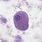
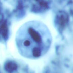
A patient who presented to the clinic is complaining of diarrhea and some abdominal discomfort. Stool specimens are submitted to the laboratory for the routine ova and parasite examination (O&P exam). When examining the permanent stained smears, the following images are seen. The test result is reported as follows: "Entamoeba histolytica trophozoites and cysts present." Based on the diagnosis of diarrhea and the laboratory findings, how would you identify the following objects seen in the permanent stained smears? Was the report correct as submitted to the physician? Why or why not?
|
|
The trophozoite on the left measures approximately 14 µm long and the cyst on the right measures approximately 13 µm. Both are stained with the standard Wheatley's modification of the Gomori trichrome, but were photographed using different filters. Remember that there is tremendous color variation using trichrome stain, both in the colors of the organisms, as well as the background debris.
Scroll Down for Answer and Discussion
Answer and Discussion of Quiz #33
Answers to Questions: The images presented above included a trophozoite and cyst of Entamoeba histolytica/E. dispar; thus the physician's report (Entamoeba histolytica trophozoites and cysts present) was incorrect. You will notice that the amebic trophozoite did not contain any ingested red blood cells (RBCs) within the cytoplasm. If no RBCs are seen in the cytoplasm and the trophozoites measure greater than or equal to 12µm, Entamoeba histolytica (true pathogen) cannot be differentiated from nonpathogenic Entamoeba dispar. Also, the cyst cannot be differentiated between the two species (E. histolytica or E. dispar) on the basis of microscopic morphology. This assumes the trophozoites are >12µm and the cysts measure >10µm. Organisms that measure below these measurements should be identified as Entamoeba hartmanni. It is also important to remember that cysts tend to shrink (more than trophozoites) after permanent staining; thus any "halo" that is seen around the cyst on the permanent stained smear must also be measured to represent the true size of the organism prior to shrinkage. Note the "halo" around the cyst at the beginning of the case history.
Currently, there are immunoassays available that will confirm the presence of organisms in the Entamoeba histolytica/E. dispar group, as well as one immunoassay that will confirm the presence of the pathogen E. histolytica.
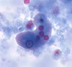
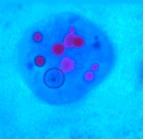
Discussion: Note that the two images seen below are trophozoites of Entamoeba histolytica, the true pathogen; this identification is confirmed by the presence of ingested RBCs within the organism cytoplasm.
The correct laboratory report for the organisms seen in Quiz #33 would have been: "Entamoeba histolytica/E. dispar trophozoites and cysts present." A computer comment could be added as follows: "Based on organism morphology, the presence of the true pathogen, Entamoeba histolytica (cause of amebiasis) could not be confirmed."
If your laboratory offers the immunoassay specific for Entamoeba histolytica, an additional computer comment could be: "If you want confirmation of the presence of Entamoeba histolytica, submit a fresh stool." Note: the immunoassays for Entamoeba histolytica or the Entamoeba histolytica/E. dispar group require fresh or frozen stools; formalinized specimens are not acceptable for testing.
|
|
Comments on the Patient: Although the patient was symptomatic, the cause may not be related to the finding of Entamoeba histolytica/E. dispar trophozoites and cysts. Since nonpathogenic intestinal protozoa, as well as pathogens, are acquired the same way through ingestion of food or water contaminated with infective cysts, this report does indicate to the physician that the patient has ingested something that was contaminated. Thus, there is always the possibility that another pathogen is involved, but was just not seen on the first fecal examination.
Comments on the Differences Between E. histolytica and E. dispar: After many years of discussion and debate, the species that is associated with amebiasis in humans is now classified as E. histolytica. The other species, which is more common but is not capable of causing invasive disease, is called E. dispar. Studies confirming the differences between the two organisms include direct sequencing of the PCR-amplified small-subunit rRNA gene of E. dispar and the design of primers for rapid differentiation from E. histolytica, differences in the phosphoglucomutases from E. histolytica and E. dispar, molecular biology of the differences in the hexokinase isoenzyme pattern, differences in the secretion of acid phosphatase, and DNA sequences unique to E. histolytica.
Clinical Disease and Therapy: Now that E. dispar has been classified as a nonpathogenic organism, totally separated from pathogenic E. histolytica, treatment is usually not recommended. However, this recommendation is based on the separation of the two species by molecular means, not by morphology alone. Since very few laboratories will be identifying the two Entamoeba species to the level of E. histolytica or E. dispar, the clinician will have to decide on the basis of clinical findings whether the patient has true pathogenic E. histolytica or nonpathogenic E. dispar infection. The laboratory report will merely indicate the presence of "Entamoeba histolytica/E. dispartrophozoites and cysts."
References:
Each Quiz has a two section format: the first section will present the Quiz topic and the second section will provide a discussion of the answer and/or various options in response to the Quiz situation presented to the user. In some situations, there may be more than one correct response.
The content within this site is made possible through the extensive contribution of Lynne S. Garcia, M.S., MT(ASCP), CLS(NCA), BLM(AAB), F(AAM), Director, Consultantation and Training Services (Diagnostic Medical Parasitology and Health Care Administration). For additional information, she can be contacted at LynneGarcia2@verizon.net.
Reference: Garcia, L.S. 2015. Diagnostic Medical Parasitology, 6th Ed., ASM Press, Washington, D.C.