

Home
About Medical Parasitology
New Infections
Ova & Parasite (O&P) Exams
CPT Codes
Quizzes, General
Quizzes, Histology
Quizzes, Blood
Review Tests
FAQ
Information Tables
Organism Index (A-Z)
***Reminder: Slides are copyrighted and cannot be copied for publication.
A 14 month old boy was admitted to the hospital with complaints of irritability, ataxia, and weakness that had developed over a period of several weeks. Just prior to admission he was unable to sit up or walk. A complete blood count showed 35% eosinophilia; lumbar puncture revealed an elevated eosinophilia. His neurologic status did not improve. Several weeks after admission his MRI revealed severe cortical atrophic changes and severe diffuse white matter degeneration. He remained in a vegetative state and died several months after his initial symptoms.
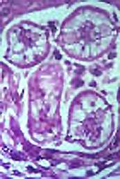
He was not seropositive for Toxocara canis or lived in or had traveled to areas where other causes of eosinophilic meningoencephalitis would be suspected. At autopsy, the following image was seen from brain tissue:
QUESTIONS
1. What are some of the other causes of eosinophilia meningoencephalitis?
2 What questions would you ask regarding possible patient history details (animal contact, play practices, etc.)?
(Scroll Down for Answers and Discussion)
ANSWER AND DISCUSSION OF DIAGNOSTIC QUIZ #59
IMAGE:
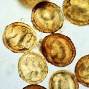
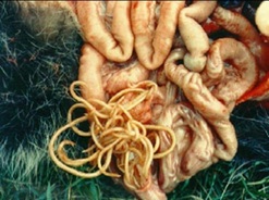
The above image reveals a histological section of larvae consistent with the raccoon roundworm, Baylisascaris procyonis. On further questioning, it became clear that the child had been exposed to feral raccoons and soil contaminated with raccoon feces in his backyard. Raccoons were seen in the surrounding area, and typical B. procyonis eggs were found in the soil (see below). In the image on the right, adult worms can be found in the raccoon.
 |
 |
 |
COMMENTS ON THE PATIENT:
The diagnosis of B. procyonis encephalitis was confirmed. This patient presented with the typical clinical syndrome, including peripheral and CSF eosinophilia. On further testing, the patient demonstrated positive antibody results with indirect immunofluorescence testing of both serum and CSF to B. procyonis. Both the intensity and titers increased with time.
COMMENTS ON BAYLISASCARIS PROCYONIS:
Feral raccoons were found in the area around the house; both dirt and plant debris were contaminated with viable B. procyonis eggs. Ingestion of infective B. procyonis eggs probably occurred through ingestion of contaminated soil or other hand-to-mouth transfer from areas or articles contaminated with raccoon feces. This roundworm is a common parasite of raccoons, especially in the Midwest, Northeast, and on the west coast of the United States. Infection rates can range as high as 82% in raccoons. In many metropolitan areas, the raccoon population is quite high, and people tend to lure the animals close to homes through intentional feeding or through dog food left in the surrounding areas. Thus, contamination of the surrounding environment with raccoon feces is common. The infected animals shed an average of >25,000 eggs/g of feces; millions of eggs can be shed each day. These infective eggs are very environmentally resistant and can survive for years in the soil.
Raccoons tend to defecate in the same areas, which are called latrines (often in barns, decks, patios, etc.). These sites remain sources of infective eggs for long periods of time for both humans and other animals. Young children with a history of ingesting soil and or feces are at greatest risk for infection; most confirmed cases have been in this age group. Individuals with developmental impairment tend to be at special risk due to a greater tendency to exhibit behaviors that lead to egg ingestion.
CLINICAL DISEASE:
Unfortunately, neural larval migrans caused by B. procyonis carries a poor prognosis. However, high-dose albendazole given early in the infection may prevent or halt the progression of CNS disease. Treatment before larvae reach the CNS is beneficial. Because they diminish inflammatory reactions, steroids may be beneficial unless the CNS damage too advanced. Although ivermectin has been used, this drug does not cross the blood-brain barrier.
In addition to direct damage caused by the migrating larvae, it is likely that eosinophil-derived neurotoxin contributes to the manifestations of encephalitis. Two features distinguish this infection from other nematode infections that cause larval migrans: the somatic migration and invasion of the CNS and the continued growth of the larvae to a large size within the CNS. Despite therapy, to date there are no documented cases of neurologically intact survivors.
EPIDEMIOLOGY:
Although reports of cases of eosinophilic meningoencephalitis caused by B. procyonis infections are rare, large populations of raccoons with high rates of B. procyonis infections live in close proximity to humans in many areas. Widespread contamination of the domestic environment suggests that the risk of exposure and human infection may be substantial. Prevention of infection is the most important public health measure; educating the public is essential for disease prevention.
REFERENCES:
Garcia, LS, 2016. Diagnostic Medical Parasitology, 6th Ed., ASM Press, Washington, DC.
Gavin, P. J., K. R. Kazacos, and S. T. Shulman. 2005. Baylisascariasis. Clin. Microbiol. Rev. 18:703-718.
Each Quiz has a two section format: the first section will present the Quiz topic and the second section will provide a discussion of the answer and/or various options in response to the Quiz situation presented to the user. In some situations, there may be more than one correct response.
The content within this site is made possible through the extensive contribution of Lynne S. Garcia, M.S., MT(ASCP), CLS(NCA), BLM(AAB), F(AAM), Director, Consultantation and Training Services (Diagnostic Medical Parasitology and Health Care Administration). For additional information, she can be contacted at LynneGarcia2@verizon.net.
Reference: Garcia, L.S. 2015. Diagnostic Medical Parasitology, 6th Ed., ASM Press, Washington, D.C.