

Home
About Medical Parasitology
New Infections
Ova & Parasite (O&P) Exams
CPT Codes
Quizzes, General
Quizzes, Histology
Quizzes, Blood
Review Tests
FAQ
Information Tables
Organism Index (A-Z)
A 9-year-old female from a rural area presented to the pediatrics outpatient department with a chief complaint of intermittent abdominal pain, anal pruritus and nocturnal restlessness since for last 2-3 weeks. On physical examination, there were no positive clinical findings. Routine O&P examinations of the stool samples revealed the following images: spherical eggs 70 µm in diameter, yellow-colored, thick-shelled eggs that contained six central hooklets but no polar filaments.
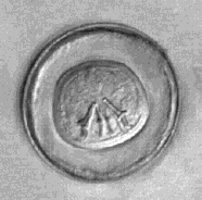
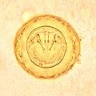

The image on the left is from the direct saline mount, while the middle and right images were prepared using D’Antoni’s iodine in the wet mount from the concentration sediment.
What organisms are suggested from the images shown above?
What groups of parasites might be involved, and are these typical based on her history?
Does the morphology provide an accurate identification? Why or why not?
How common is this parasite worldwide and how likely is it that the patient might be asymptomatic?
The images certainly suggest a helminth egg; the overall appearance is that of a tapeworm egg (cestode), particularly with the presence of the six-hooked embryo (oncosphere).
The history is relatively non-specific; in cases of this infection there may be nothing to suggest the infection and/or possible exposure route. Also, the patient may actually be asymptomatic.
The presence of spherical eggs 70 µm in diameter, yellow-colored, thick-shelled eggs that contained six central hooklets but no polar filaments are suggestive of Hymenolepis diminuta eggs, the rat tapeworm. Although Hymenolepis nana (the dwarf tapeworm) is much more common, occasionally
H. diminuta can be found, particularly in children. The H. diminuta eggs are characterized by having no polar filaments that lie between the oncosphere and the egg shell, while H. nana has the typical polar filaments (see images below).
This infection is uncommon, but when found is often from children. Again, the patient may have vague mild symptoms or may be asymptomatic.
COMMENTS ON THE PATIENT and INFECTION:
H. diminuta is a rodent parasite for which the arthropod acts as intermediate host. Rodents become infected by ingesting the arthropod containing cysticercoid larvae. Human beings, usually children, can be accidentally infected by the ingestion of infected arthropod (often grain beetles). In humans, infections with
H. nana are much more common than those by H. diminuta, as its transmission does not require any intermediate host. H. nana can be spread directly from person to person via the eggs. Although H. diminuta infection in humans is uncommon, a few hundred cases have been reported worldwide. The human form of H. diminuta infection is often asymptomatic, but abdominal pain, irritability, itching and eosinophilia have been reported.
COMMENTS ON THE METHOD RECOMMENDATIONS:
The routine O&P examination is recommended for egg recovery and subsequent microscopic identification (10X and 40X power – low and high dry).
COMMENTS ON THE IMAGES:
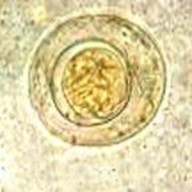
The images are very typical. Note the six-hooked oncosphere and no polar filaments in H. diminuta, while the polar filaments are present in H. nana (see arrows).

Hymenolepis diminuta Hymenolepis nana
Garcia, L.S. 2016. Diagnostic Medical Parasitology, 6th Ed., ASM Press, Washington, D.C.
Each Quiz has a two section format: the first section will present the Quiz topic and the second section will provide a discussion of the answer and/or various options in response to the Quiz situation presented to the user. In some situations, there may be more than one correct response.
The content within this site is made possible through the extensive contribution of Lynne S. Garcia, M.S., MT(ASCP), CLS(NCA), BLM(AAB), F(AAM), Director, Consultantation and Training Services (Diagnostic Medical Parasitology and Health Care Administration). For additional information, she can be contacted at LynneGarcia2@verizon.net.
Reference: Garcia, L.S. 2015. Diagnostic Medical Parasitology, 6th Ed., ASM Press, Washington, D.C.