

Home
About Medical Parasitology
New Infections
Ova & Parasite (O&P) Exams
CPT Codes
Quizzes, General
Quizzes, Histology
Quizzes, Blood
Review Tests
FAQ
Information Tables
Organism Index (A-Z)
Gnathostomiasis, Gnathostoma spinigerum, G. doloresi, G. nipponicum, G. hispidum, G binucleatum (Pathogen - Tissue Nematode)
Organism:
Human gnathostomiasis is an emerging food-borne parasitic disease caused by nematodes in the genus Gnathostoma. Human gnathostomiasis caused by Gnathostoma spp. is a highly endemic disease in some under-developed communities in Asia, particularly in China. Recently, it has become an emerging disease among travelers from Europe and other continents who are coming into contact with endemic areas. Nematodes of the genus Gnathostoma (Nematoda: Gnathostomatidae) are the etiological agents of human gnathostomiasis and may also infest dogs, cats, tigers and leopards(3). There are 12 species in this genus with four species recorded in humans: G. spinigerum, commonly found in India, China, Japan and Southeast Asia; G. hispidum, found in Europe, Asia and Australia; G. doloresi, found in Southeast Asia; and G. nipponicum, found in Japan. G. spinigerum is frequently reported in Asia as being responsible for human gnathostomiasis, but G. hispidum, G. doloresi, and G. nipponicum have also occasionally been reported.
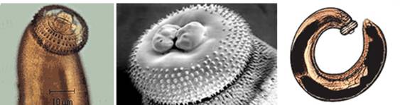
Left, Third-stage larva of Gnathostoma spinigerum; Center, scanning electron micrograph of a female Gnathostoma spinigerum head bulb; Right, whole larva of Gnathostoma spinigerum (Courtesy of the CDC Public Health Image Library).


![]()
![]()
Left, Example of cutaneous gnathostomiasis; Right, Examples of ocular gnathostomiasis (Courtesy of Dr. J. Baquera).
Life Cycle:
Man is an accidental host and represents the dead end for the parasite. Human infection is mainly resulted from eating raw or undercooked intermediate hosts (e.g. fish, eels, and loaches) or paratenic hosts (e.g. crustaceans, freshwater fish, and mammals) containing the third-stage (L3) larvae. The larvae cannot mature in humans and keep migrating in the skin, subcutaneous tissues, or other organs. Gnathostomiasis is characterized by intermittent creeping eruptions and/or migrating swellings and eosinophilia; larval migration to other tissues (visceral larva migrans) can result in a serious consequence. If untreated, gnathostomiasis may remit and recur several times until death of the larvae up to 10+ years after infection. Although the parasite fails to reach sexual maturity in humans, they may remain alive up to 10 years
Acquired:
Human infection is acquired from eating raw or undercooked intermediate hosts (e.g. fish, eels, and loaches) or paratenic hosts (e.g. crustaceans, freshwater fish, and mammals) containing the third-stage (L3) larvae.
Epidemiology:
The endemic foci of gnathostomiasis have been predominantly distributed in Japan and Southeast Asia, particularly Thailand, but the disease is also endemic in Cambodia, Laos, Myanmar, Indonesia, the Philippines, and Malaysia. Human cases have also been reported in India, Australia, Brazil, and parts of South Africa, and it has been regarded as an emerging disease. Imported human cases of gnathostomiasis were also reported in the Republic of Korea. Recently, it has been known that this disease is endemic in the Pacific region of Mexico. In China, gnathostomiasis occurred sporadically in 23 Provinces, Autonomous regions, or Municipalities.
In parts of Asia, wild-caught and aquaculture-reared swamp eels (Synbranchidae: Monopterus spp.) are widely consumed as food by humans and are a common source of human gnathostomiasis, a foodborne zoonosis caused by advanced third-stage larvae (AL3) of Gnathostoma spp. nematodes. Over the past 2 decades, many thousands of swamp eels (Synbranchidae: Monopterus spp.) have been legally shipped alive from Asia to North America, where they were distributed to numerous ethnic food markets in major cities in the United States and Canada. An earlier survey of live Asian swamp eels from ethnic markets in the United States and introduced wild populations in Florida found substantial parasite burden in both market and wild swamp eels sampled; however, the researchers did not examine eels for Gnathostoma spp.
Clinical Features:
Gnathostomiasis can be divided into cutaneous, visceral, and ocular forms, depending on the site of larval migration and subsequent signs and symptoms. The most severe manifestation of the visceral disease is involvement of the central nervous system. Gnathostoma spp. nematodes are highly invasive parasites. After being ingested, the third-stage larvae penetrate the mucosal wall of the gastrointestinal tract. Larvae can enter the CNS by invading directly through the loose connective tissues of the neural foramina of the skull base along the cranial nerves and vessels or through intervertebral foramina along the spinal nerves and vessels.
Gnathostomiasis is an important food-borne helminth infection, with reports primarily from Thailand, Japan, and other Southeast Asian countries.