

Home
About Medical Parasitology
New Infections
Ova & Parasite (O&P) Exams
CPT Codes
Quizzes, General
Quizzes, Histology
Quizzes, Blood
Review Tests
FAQ
Information Tables
Organism Index (A-Z)
Dipylidium caninum (Pathogen – Intestinal Cestode)
Organism:
D. caninum is commonly found throughout the world in dogs and cats, both domestic and wild. Human infections have also been reported from many areas of the world, including the United States. Most reported infections have been in children.
 |
 |
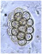 |
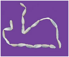
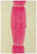
| D. caninum strobila of proglottids | D. caninum proglottid | D. caninum egg packet |
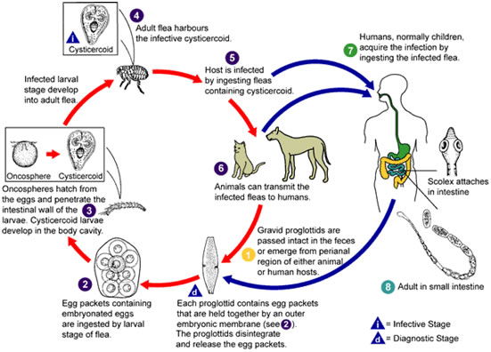
Life Cycle:
The life cycle is very similar to that of H. diminuta, in which the arthropod is an obligatory intermediate host. The adult worms are found in the dog or cat intestine, and gravid proglottids separate from the strobila and may migrate singly or in short chains out of the anus. The eggs are ingested by the larval stages of the dog, cat, or human flea, where they develop into cysticercoid larvae. When these fleas are then ingested by the definitive host (dogs, cats, or humans), the adult worm develops within 3 to 4 weeks.
Acquired:
Infection in humans is acquired through ingestion of immature tapeworm larvae, cysticercoids, from infected arthropods.
Epidemiology:
Worldwide, primarily human to human transmission
Clinical Features:
The symptoms are related to worm burden; however, in most patients (usually children), some complaints may consist of indigestion and appetite loss. Awareness of the infection may first appear from the migration of proglottids from the anus. Most human cases have been in children, indicating that children may be more likely to accidentally swallow the infected fleas or may be more susceptible to infection. Periodic administration of anthelmintic agents to dogs and cats and use of flea powders will help reduce the risk of infection.
Clinical Specimen:
Stool: The standard O&P examination is the recommended procedure for recovery and identification of D. caninum egg packets in stool specimens, primarily from the wet preparation examination of the concentration sediment.
Adult worms: Gravid proglottids may be recovered in stool; often they can be seen lying on the top or bottom of the stool specimen submitted as a fresh specimen. Often a partial chain of proglottids may be passed (a few inches to several feet).
Laboratory Diagnosis:
Stool: The standard O&P examination is the recommended procedure for recovery and identification of D. caninum eggs in stool specimens, primarily from the wet preparation examination of the concentration sediment. The eggs are most easily seen on a direct wet smear or a wet preparation of the concentration sediment.
Adult worms The mature and gravid proglottids are wider than long, with the main reproductive structures (mainly the uterus) located in the center of the gravid proglottid. This configuration of the uterine structure has been called a rosette.
Organism Description:
Egg: Groups of eggs (egg packets) may be found in the stool. Each egg measures from 25 to 40 µm and contains the six‑hooked oncosphere. The individual eggs may closely resemble those of Taenia spp., particularly if they are released from the egg packet.
Adult worm: The adult worms measure from 10 to 70 cm long and have a scolex with four suckers and an armed rostellum. The single proglottids have been described as looking like cucumber seeds when moist and like rice grains when dry.
Laboratory Report:
Dipylidium caninum eggs present
Dipylidium caninum proglottids.
Treatment:
Garcia, L.S. 2007. Diagnostic Medical Parasitology, 5th ed., ASM Press, Washington, D.C.
Control:
This infection is a true zoonosis, since infected dogs and cats infect fleas, which in turn are accidentally consumed by humans. Children may be more likely to accidentally swallow the infected fleas or may be more susceptible to infection. Periodic administration of anthelmintic agents to dogs and cats and use of flea powders will help reduce the risk of infection.