

Home
About Medical Parasitology
New Infections
Ova & Parasite (O&P) Exams
CPT Codes
Quizzes, General
Quizzes, Histology
Quizzes, Blood
Review Tests
FAQ
Information Tables
Organism Index (A-Z)
Plasmodium vivax (True Pathogen –Tertian Malaria)
Organism:
This organism belongs to the phylum Apicomplexa, is a true pathogen, and causes malaria. All forms of the human life cycle will appear in the thick and thin blood films.
 |
 |
 |
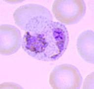
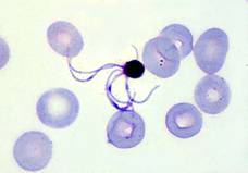
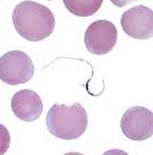
Plasmodium vivax ring forms schizont NOTE: Enlarged RBC's, ameboid rings, stippling |
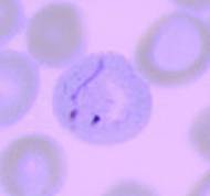
Plasmodium vivax mature Thick Blood Film |
Life Cycle:
Within an hour of infection, sporozoites from the mosquito are carried via the blood to the liver, where they penetrate parenchymal cells, thus initiating the preerythrocytic or primary exoerythrocytic cycle. The sporozoites become round or oval and begin dividing, resulting in large numbers of liver merozoites. The merozoites leave the liver and invade the red blood cells (RBCs), initiating the erythrocytic cycle. In P. vivax and P. ovale, a secondary or dormant schizogony occurs from organisms that remain quiescent in the liver until a later time; they are called hypnozoites. Delayed schizogony does not occur in P. falciparum, P. malaria, or P. knowlesi.
P. vivax infects only the reticulocytes and the parasitemia is usually limited to 2 to 4% of the available RBCs. Splenomegaly occurs during the first few weeks, and the spleen will progress from being soft and palpable to hard during a chronic infection. If therapy is given early, the spleen will return to normal size. Leukopenia is seen; leukocytosis may be present during the febrile episodes. Total plasma proteins are unchanged, although the albumin may be low and the globulin fraction may be elevated due to antibody development. Serum potassium may also be increased.
Acquired:
Bite: female anopheline mosquito; blood, shared needles, congenital infections
Epidemiology:
P. vivax may account for 80% of the infections; tropics, subtropics, and temperate zones, mosquito to human, human to human transmission
Clinical Features:
Early Infection: The primary clinical attack usually occurs from 7 to 10 days after infection, although there are strain differences, with a much longer incubation period being possible. In some patients, symptoms such as headache, photophobia, muscle aches, anorexia, nausea, and sometimes vomiting may occur before organisms can be detected in the bloodstream. In other patients, the parasites can be found in the bloodstream several days before symptoms appear. During the first few days, the patient may not exhibit a typical paroxysm pattern but rather have a steady low‑grade fever or an irregular remittent fever pattern. Once the typical paroxysms begin, after an irregular periodicity, a regular 48‑h cycle is established. An untreated primary attack may last from 3 weeks to 2 months or longer. The paroxysms become less severe and more irregular in frequency and then stop altogether. In 50% of the cases, relapses may occur after weeks, months, or up to 5 or more years.
Complications: Severe complications are less common in P. vivax infections, although coma and sudden death or other symptoms of cerebral involvement have been reported. Severe sequelae can be seen in cases of primaquine-tolerant or primaquine-resistant cases.
Clinical Specimen:
Blood: Multiple draws (EDTA). Malaria is one of the few parasitic infections considered to be immediately life‑threatening, and a patient with the diagnosis of P. falciparum malaria should be considered a medical emergency because the disease can be rapidly fatal. Any laboratory providing the expertise to identify malarial parasites should do so on a 24‑h basis, 7 days/week. Prepare thick and thin blood films immediately after receipt of the blood.
Laboratory Diagnosis:
Although malaria is no longer endemic within the United States, this infection is life threatening, and laboratory requests for blood smear examination and organism identification should be treated as “STAT” requests. Frequently, for a number of different reasons, organism recovery and identification may be more difficult than the textbooks imply. It is very important that this fact be recognized, particularly when one is dealing with a possibly fatal infection with P. falciparum or P. knowlesi. Both thick and thin blood films should be prepared on admission of the patient (clinic, emergency room, in-house), and at least 300 oil immersion fields should be examined on the thick and thin film before a negative report is issued. Since one set of negative films will not rule out malaria, additional blood specimens should be examined over a 36 h time frame. Although Giemsa stain is used for parasitic blood work; the organisms can also be seen with other blood stains such as Wright’s, Wright-Giemsa, or any of the rapid blood stains. Blood collected with EDTA anticoagulant is acceptable; however, if the blood remains in the tube for any length of time, true stippling may not be visible within the infected RBCs (P. vivax as an example), organisms will change their morphology, and some of the parasites will disintegrate (after 4-6 h). Also, it is important to remember that the proper ratio between blood and anticoagulant is necessary for good organism morphology.
Organism Description:
Ring Forms: Ring forms of P. vivax tend to be delicate, spread out (ameboid), and may mimic ring forms of the other species. Stippling (Schüffner’s dots) do not appear immediately as they do in P. ovale, but appear a few hours later. However, if the blood in EDTA stands for >one hour, the stippling may not be seen, regardless of the pH of the buffer.
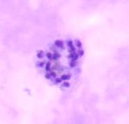
Mature Schizont : Often quite large containing 18+ merozoites.
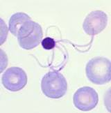
Gametocyte: The gametocytes are round and tend to fill the entire RBC. Exflagellation of the male gametocytes can occur in the blood if the blood cools and becomes aerated (cap removed – lag time between cooling/cap removal and preparation of the thick and thin blood films.
Laboratory Report:
A number of reports can be relevant – remember to add the appropriate report comments.
Report 1: No Parasites Seen: The submission of a single blood specimen will not rule out malaria; submit additional bloods every 4-6 hours for 3 days if malaria remains a consideration.
Report 2: Plasmodium spp. Seen: Unable to rule out Plasmodium falciparum or Plasmodium knowlesi
Report 3: Plasmodium spp., possible mixed infection: Unable to rule out Plasmodium falciparum or Plasmodium knowlesi
Report 4: Negative for parasites using automated hematology instruments. Automated Hematology instruments will not detect low malaria parasitemias seen in immunologically naïve patients (travelers)
Treatment:
Garcia, L.S. 2007. Diagnostic Medical Parasitology, 5th ed., ASM Press, Washington, D.C.
Control:
Improved mosquito control, impregnated bed nets, potential vaccines, prophylaxis
Comments:
It is important to remember that the malarial parasites remain viable in the tube of blood collected using EDTA. If the blood cools and the cap is removed (blood becomes aerated), the organisms then begin the life cycle seen in the mosquito. Thus exflagellation of the male gametocyte can mimic spirochetes when they separate from the gametocyte (see below):
 |
 |
 |