

Home
About Medical Parasitology
New Infections
Ova & Parasite (O&P) Exams
CPT Codes
Quizzes, General
Quizzes, Histology
Quizzes, Blood
Review Tests
FAQ
Information Tables
Organism Index (A-Z)
Dracunculiasis, Dracunculus medinensis (Pathogen - Tissue Nematode)
Organism:
The contemporary term, guinea worm disease, derives its name from a European explorer who named the disease for the geographic area in which it was found, along the western African coast. The staff of Aesculapius, Roman god of medicine, may have originated from the ancient, still used procedure of removing the adult worm by slowly winding it around a stick. Although the worms are very long and thin, they are not true filarial worms but, rather, are grouped in their own order within the nematodes. Most countries, including Asia, are declared free from the Guinea worm disease; thus, the burden of transmission remains in Africa, especially Chad, Ethiopia, Mali, and South Sudan.
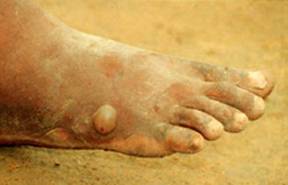
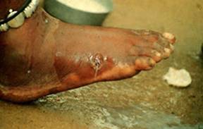
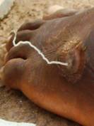
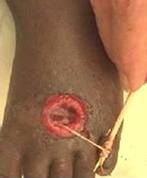
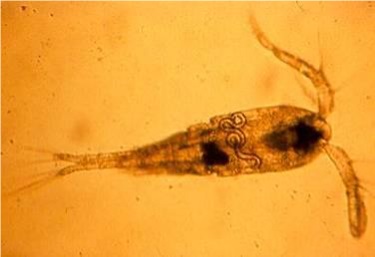
Upper Left, Dracunculus medinensis blister; Upper Right, open blister with evidence of the worm; Middle Left, worm coming out of the ulcerated blister; Middle Right, worm removal by winding the worm onto the stick; Bottom, Copepod with D. medinensis larva inside (Courtesy of the WHO Collaborating Center at CDC archives.
Life Cycle:
Human infection is acquired from ingestion of infected copepods (Cyclops water fleas). The released larvae penetrate the duodenal mucosa and develop in the loose connective tissue. The possibility also exists that paratenic hosts, such as tadpoles and frogs, are important means of transporting infective larvae of Dracunculus species up the food chain, thus facilitating transmission to the definitive hosts. The worms mature in the deep connective tissue, and the females migrate to the subcutaneous tissues when they are gravid and contain coiled uteri filled with rhabditiform larvae. Maturation takes approximately 1 year. At this stage in the life cycle, the female migrates to the skin and a papule is formed in the dermis, usually by the ankles or feet (although papules can be anywhere on the body). The papule changes into a blister within 24 h to several days. Eventually, the blister ulcerates, and on contact with freshwater, a portion of the uterus prolapses through the worm’s body wall, bursts open, and discharges thousands of larvae into the water. This may happen several times until all of the larvae are discharged. The larvae are then ingested by an appropriate species of Cyclops. Development takes about 8 days before the larvae are infective for humans.
Acquired:
Human infection is acquired from ingestion of infected copepods (Cyclops water fleas)
Epidemiology:
Disease transmission depends on several factors: (i) water sources where Cyclops spp. breed, (ii) direct contact between infected humans and the water source, (iii) use of this water source for drinking, or (iv) the possibility of other paratenic hosts. In various parts of the world, certain types of water sources (e.g., step wells in India, covered cisterns in Iran, and ponds in Ghana) provide all of these transmission requirements. The disease can be eliminated within 1 to 2 years by provision of safe drinking water.
Clinical Features:
After ingestion of an infected copepod, no specific pathologic changes are associated with larval penetration into the deep connective tissues and maturation of the worms. Once the gravid female begins to migrate to the skin, there may be some erythema and tenderness in the area where the blister will form. Several hours before blister formation, the patient may exhibit some systemic reactions, including an urticarial rash, intense pruritus, nausea, vomiting, diarrhea, or asthmatic attacks. The lesion develops as a reddish papule, measuring 2 to 7 cm in diameter. Symptoms usually subside when the lesion ruptures, discharging both the larvae and worm metabolites.
Clinical Specimen:
Portions of the adult worm(s) may be identified.
Laboratory Diagnosis:
Diagnosis can be confirmed at the time the cutaneous lesion forms, with subsequent appearance of the adult worm. Infected lesions must be distinguished from carbuncles, deep cellulitis, focal myositis or periostitis, and even rheumatism. Calcified worms may also be found in subcutaneous tissues by radiography. They may appear as linear densities (up to 25 cm), tightly coiled structures, or sometimes nodules. Depending on the site, they can also be misdiagnosed as possible breast cancer.
Organism Description:
The worms are very long, with the females measuring up to 1 m in length by 2 mm in width. The male is much smaller and inconspicuous (2 cm long).
Treatment:
For centuries, the worms have been removed by slowly being wound around a stick. This approach works well unless the worm is accidentally broken and secondary infection occurs. Allergic manifestations can be decreased by using epinephrine. Four drugs have been used with various degrees of success: niridazole, thiabendazole, metronidazole, and mebendazole. The action seems to involve suppression of inflammation rather than any specific effect on the adult worms, although 400 to 500 mg/day for 6 days has been reported to kill the worms directly. The prognosis is usually quite good unless there are complications such as chronic recurrent nodules and ulcers, aberrant migration of the worms, or calcification of the adult worms.
Control:
Through January of 2013 Asia was free from dracunculiasis, and no cases were reported worldwide for the first time since the eradication program began in 1986. It is hoped that this infection will eventually join smallpox as one that can actually be eradicated from the world.