

Home
About Medical Parasitology
New Infections
Ova & Parasite (O&P) Exams
CPT Codes
Quizzes, General
Quizzes, Histology
Quizzes, Blood
Review Tests
FAQ
Information Tables
Organism Index (A-Z)
The microsporidia are obligate intracellular parasites that have been recognized in a variety of animals, particularly vertebrates. Typical sizes of the spores range from 1.5 to 2.0um in humans. The infectious stage, the spore, contains a coiled polar tubule, which is an extrusion mechanism for injecting the infective spore contents into host cells. Demonstration of the coiled polar tubule within spores is diagnostic for microsporidial infections. Currently, there are eight genera of microsporidia that can infect humans. the more common are Encephalitozoon spp., Septata intestinalis and Enterocytozoon bieneusi; the less common are Brachiola spp, Microsporidium spp., Nosema spp., Pleistophora spp., and Trachipleistophora spp., and Vittaforma spp.. Essentially every body site can be infected with one or more of these eight genera; some organisms will tend to disseminate to other parts of the body from the primary site of intection (often the GI tract).
Human infections occur through ingestion, inhalation, and probably direct inoculation of infectious spores from the environment. Infection occurs with the introduction of infective sporoplasm through the polar tubule into the host cell. Microsporidia multiply extensively with the host cell cytoplasm; the life cycle includes repeated divisions by binary fission (merogony) or multiple fission (schizogony) and spore production (sporogony). During sporogony, a thick spore wall is formed, providing environmental protection for this infectious stage of the parasite.
Conjunctival and corneal epithelium infections occur in HIV patients, while corneal stroma and ulceration occur in immunocompetent individuals. The chronic intractable diarrhea, fever, malaise, and weight loss of E. bieneusi infections are similar to symptoms of cryptosporidiosis or isosporiasis. These patients have been diagnosed with AIDS and they tend to have 4 to 8 watery, non-bloody stools per day, which can be accompanied by nausea and anorexia. There may also be dehydration and fat malabsorption. These patients tend to be severely immunodeficient, with a CD4+ count always below 200 and often below 100. Concurrent infections with Encephalitozoon intestinalis have been reported.
Unlike E. intestinalis, E. bieneusi infections do not respond to albendazole therapy. For the most part, therapy for microsporidial infections is difficult, at best.
The standard O&P concentration is recommended (500 x g for 10 min) with the concentrate sediment being used for all smears and for the FA procedure. Recommended stains include the modified trichrome stains (Weber Green modified trichrome; Ryan Blue modified trichrome). Stool preparations must be very thin, the staining time is 90 min, and the slide must be examined at X1,000 magnification. Unfortunately, stool material contains a lot of artifacts, many of which will mimic microsporidian spores. If the modified trichrome stains are used for stool specimens, positive control material should be available for comparison. There are other stains available, some including heat and some being combined with modified acid-fast stains. Another approach involves chemofluorescence using optical brightening agents (Calcofluor White, Fungi-Fluor, or Uvitex 2B); these reagents are sensitive, but nonspecific. This is a problem when examining stool specimens; both false positive and false negative results have been seen. Currently, there are no commercially available immunoassay reagent kits available. Examination of other body site specimens (sputum, joint fluids, eye specimens, etc.) is likely to be easier than looking at stool specimens; spores are not as likely to be confused with artifacts.
Histologic diagnosis can be accomplished using routine hematoxylin and eosin stains; however, modified tissue Gram stains, PAS, and silver stains are also recommended. Smear preparations (eye, etc.) can also be stained using Giemsa stain.
Little information is available regarding reservoir animal hosts for human microsporidia or environmental survival times; however, the spores tend to be very resistant to adverse conditions and can survive for long periods of time if kept moist. There is every reason to believe that the spores can be transmitted through contaminated food and/or water.
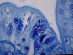
This is a routine histologic preparation of intestinal tract tissue stained with Giemsa stain. Note the small dots within some of the cells. These are microsporidian spores undergoing development. Each spore measures about 1-1.5 μm. The intestinal cells will slough off and pass out in the stool; often free spores can be seen in the stool specimen after processing and special staining with modified trichrome stains.
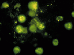
This photograph shows a urine sediment specimen stained with an experimental FA reagent. Note the oval spores within some of the urine sediment cells. There are also a couple of spores outside of the cells. These experimental reagents are specific for microsporidia in the genus, Encephalitozoon spp.; the kidney infection may represent dissemination from the GI tract.
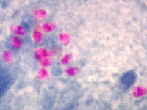
This is a fecal specimen stained with the Ryan Modified Trichrome Stain. The pink structures are the microsporidian spores that will measure approximately 1-2μm. They will appear a bit larger in the stool preparation than they will within the tissue. However, this photograph has also been enlarged to show the key characteristic, the polar tubule (polar filament). Note that some of the spores appear to have a horizontal line across the middle of the spore; this is evidence of the polar tubule. The specimen was taken from concentration sediment (centrifugation 10 min at 500 Xg).
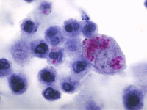
This preparation is also urine sediment stained with Giemsa stain. Note the small pink spores within the cells in the sediment. Again, when inside the cells, they will appear somewhat smaller than they will outside of the cells in the background.